
There is no single sleeping heart-rate range that is normal for every person. In many people, heart rate is lower during non-REM sleep than during calm wakefulness. It usually becomes faster and more variable during REM sleep, and it can rise briefly with an arousal. The meaning of any nighttime number depends on age, personal baseline, fitness, pregnancy, illness, medicines and substances, sleep stage, heart rhythm and how the measurement was made.
The familiar adult resting range is not a sleeping target. The American Heart Association gives 60 to 100 beats per minute as a resting reference for most adults who are sitting or lying down, calm and feeling well. It also notes that heart rate may fall below 60 during sleep 1. That does not establish 40 to 60, or any other interval, as a universal healthy sleeping range.
A single average or minimum from a watch cannot show whether a heart rhythm is normal. Look at the person, the pattern and the source of the measurement before deciding what the number means.
What normally happens to heart rate during sleep
The nervous system changes how it regulates the heart as sleep progresses:
- Non-REM sleep: Heart rate and blood pressure generally fall below relaxed waking levels. The pattern is usually more stable, especially in deeper non-REM sleep.
- REM sleep: Average cardiovascular activity and beat-to-beat variability increase. Short accelerations and irregular-looking changes can occur during the more active parts of REM.
- Arousals and awakenings: A brief shift toward wakefulness can produce a short rise in heart rate, even when the person does not remember waking.
These stage-related patterns are established physiology, but their size varies between people and across the night 2. There is no reliable rule that everyone’s heart rate must fall by a certain percentage after sleep begins.
A nightly average blends different stages, positions and events. A minimum may represent only a short period. A graph with minute-by-minute values provides more context than either number alone, but it still cannot identify the electrical rhythm unless the device recorded a usable ECG.
Heart rate, heart rhythm and pulse are not the same question
Heart rate is the number of heartbeats per minute. Heart rhythm describes where the electrical impulses begin and whether the beats follow an expected sequence. A rate of 65 beats per minute, for example, can occur with different rhythms.
A manual wrist pulse and most overnight wearables do not directly count the heart’s electrical signals. They detect pressure or blood-volume pulses that reach the skin. Under good conditions, pulse rate is a useful estimate of heart rate. With an irregular rhythm, weak peripheral pulse, poor sensor contact or artifact, the pulse estimate and electrical heart rate may not match 3.
This distinction explains why a normal-looking average cannot rule out an arrhythmia and an isolated extreme watch value cannot diagnose one. Rhythm questions require an ECG-quality recording interpreted in clinical context.
Why personal baseline matters
Resting heart rate differs considerably between people. In a longitudinal study of more than 92,000 adults using wrist-worn trackers, each person’s daily resting rate was generally more consistent than the range seen across the full population. Age, sex, body size, sleep duration and season explained only part of the between-person variation 4.
That study used a device-derived daily resting metric, not a clinical definition of normal sleeping heart rate. Its practical lesson is still useful: a repeated change from a stable personal pattern can be more informative than comparing one night with a generic internet range.
Compare like with like. A watch’s nightly average should be compared with prior nights from the same device and similar wear conditions, not with a spot pulse, a sleep-lab ECG minimum or another brand’s proprietary metric.
Adults and children need separate interpretation
Adult cutoffs should not be applied to infants or children. Heart rate is generally faster earlier in childhood and changes with development. The relevant reference also depends on whether the child is awake, asleep, ill, distressed or taking a medicine that affects rate.
Even pediatric sleep data do not support one all-age table. A study of two cohorts of healthy children ages 6 through 11 found that sleeping heart rate decreased with age and differed with sex, body size and cohort. One cohort used full polysomnography, while the other could have included quiet wakefulness in its average 5. Those findings cannot define a normal value for an infant, teenager or individual child.
Ask a pediatric clinician to interpret a persistent or symptomatic change using the child’s exact age, clinical condition and measurement method. Seek urgent care for severe symptoms rather than waiting to compare the number with a chart.
What can change sleeping heart rate
A different nighttime number is not automatically a heart disorder. Common contexts include:
Sleep stage, movement and arousal
REM periods, turning over, waking briefly, pain, a nightmare or an environmental disturbance can produce short increases. A smoother section during non-REM sleep and short peaks around arousals can be physiological. A consumer wearable cannot reliably assign the cause of each peak.
Breathing events
Obstructive or central breathing events can be accompanied by oxygen changes, arousal and shifts in autonomic activity. Sleep-disordered breathing is also associated with several bradyarrhythmias and tachyarrhythmias, but a heart-rate trace alone cannot diagnose sleep apnea or establish that a breathing event caused a spike 6.
Discuss repeated changes with a clinician when they occur alongside loud snoring, witnessed pauses, gasping, marked daytime sleepiness or an existing heart condition. Do not start, stop or change PAP treatment based on a wearable heart-rate graph.
Fever, illness, pain and recovery
Body temperature, pain and emotional stress can raise heart rate. Vomiting, diarrhea, reduced intake and other illness-related changes can also alter the nightly baseline. The AHA advises interpreting heart rate in the context of temperature, emotions, body position and medicines rather than as an isolated number 1.
A temporary rise during illness may settle as the illness improves. Persistent changes, concerning symptoms or a high-risk medical history warrant clinical advice.
Alcohol, stimulants and other substances
Alcohol may feel sedating while still raising nighttime heart rate. In a small randomized sleep-laboratory study of healthy adults ages 30 to 60, presleep alcohol increased nocturnal heart rate in a dose- and time-dependent pattern compared with placebo 7. The study does not predict the exact response for every person or establish a safe bedtime dose.
Caffeine, nicotine, cannabis, decongestants and other substances may also affect rate, rhythm or sleep 89. Record what was used and when. Do not use a change in the watch graph as a reason to abruptly stop a prescribed medicine.
Medicines and medication changes
Beta blockers, some calcium-channel blockers and several other medicines can slow heart rate. Other prescriptions and over-the-counter products can raise it or affect rhythm. The relevant question is whether the observed pattern is expected for the specific medicine, dose, timing, condition and person 19.
Contact the prescriber when a new pattern follows a start, stop or dose change, especially if there is weakness, dizziness, near-fainting, breathlessness or palpitations. Do not adjust the medicine on your own.
Fitness and physical conditioning
Physical conditioning can contribute to a lower resting baseline. It does not prove that every low nighttime rate in an active person is harmless. Symptoms, rhythm, medicines, sleep apnea, medical history and the reliability of the reading still matter.
Cardiology guidance emphasizes matching symptoms to documented bradycardia and evaluating reversible or physiological causes. It also recommends sleep-apnea assessment when sleep-related bradycardia or a conduction disorder raises clinical suspicion 9.
Pregnancy
Maternal heart rate changes across pregnancy, so a prepregnancy baseline may not remain the right comparison. A systematic review found that heart rate rose across gestation on average, with substantial differences between studies and insufficient evidence for a precise gestation-specific normal range 10.
Pregnancy-specific symptoms matter. Promptly contact maternity care for a new persistent racing or irregular heartbeat, especially with dizziness, fainting, chest symptoms or breathing difficulty. Severe symptoms need emergency care.
An arrhythmia
A rhythm problem may be fast, slow or irregular, and it may be intermittent. Some produce palpitations, weakness, dizziness, fainting, breathlessness or chest discomfort; others cause no obvious symptoms. Neither a normal nightly average nor a single watch alert can settle the diagnosis.
How accurate is a smartwatch during sleep?
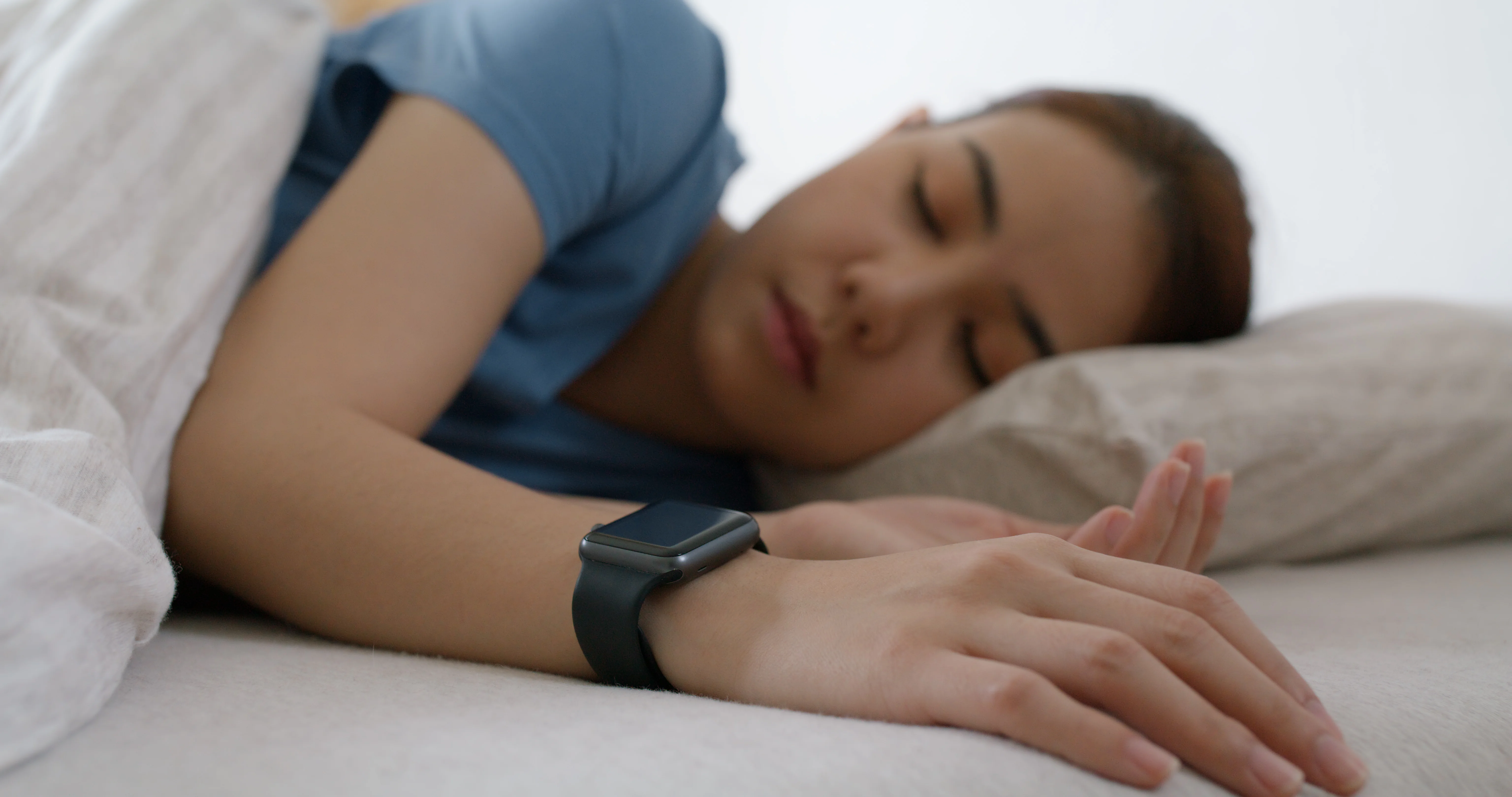
Most watches and rings use photoplethysmography, or PPG. LEDs shine light into the skin and a sensor detects pulse-related changes in reflected light. An algorithm turns those pulse waves into a rate estimate.
PPG is useful for trends, especially when the signal is clean, but it is not the same as ECG. Relevant limitations include:
- loose or inconsistent skin contact
- movement, pressure on the sensor or sleeping on the wrist
- low peripheral blood flow or cold skin
- tattoos, hair and other local optical factors
- irregular rhythms and weak pulse waves
- filtering, averaging and proprietary device algorithms
- differences between device models and software versions
Evidence about skin pigmentation and wrist-wearable heart-rate accuracy is mixed. A systematic review found reduced accuracy with darker skin in some studies, no difference in others and mixed device-specific findings in the remainder. The included studies were small and heterogeneous, so neither universal bias nor universal equivalence has been established 11.
Treat an isolated implausible point as something to verify, not something to ignore or diagnose. Check whether the device was fitted as directed, whether the graph has a signal gap and whether the finding repeats.
Which measurement answers which question?
A manual spot pulse
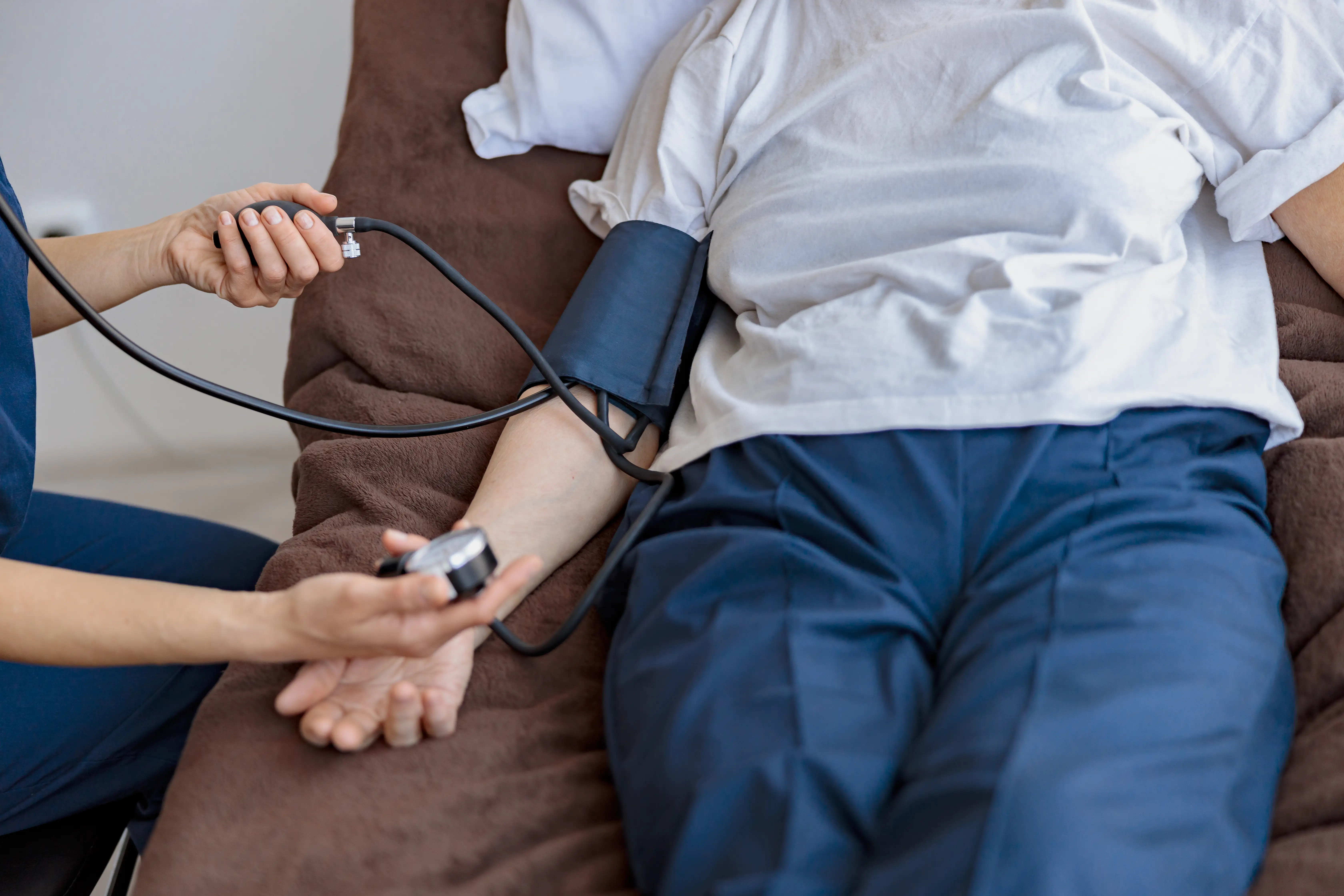
A pulse check while awake can confirm the approximate current rate and whether the pulse feels regular. The AHA describes counting the wrist pulse for a full 60 seconds 1. This check cannot reconstruct what happened during sleep, and an irregular or weak pulse needs clinical assessment rather than repeated counting.
A standard ECG
A clinical ECG records the heart’s electrical activity and is the most common test used to diagnose an arrhythmia. It shows the rhythm during a short recording window. A normal ECG can therefore miss an intermittent event that happened overnight 12.
Ambulatory ECG monitoring
A Holter monitor, patch, event monitor or loop recorder records electrical rhythm over a longer period. Clinicians choose the monitor according to how often the event occurs and what they need to capture. Longer monitoring can link a symptom or nighttime event to the actual rhythm when a brief office ECG does not 12.
Polysomnography
An attended sleep study records sleep stage, breathing, oxygen and a single ECG channel together. The report can show whether a rate or rhythm change occurred during wake, non-REM or REM sleep and whether it coincided with a respiratory event or oxygen change 13.
Polysomnography is useful when the clinical question involves sleep or sleep-disordered breathing. Its standard single ECG channel is not a substitute for a full diagnostic ECG or appropriately selected ambulatory rhythm monitor.
A practical response to an unusual nighttime reading
1. Start with symptoms
Severe symptoms determine urgency. Do not spend time troubleshooting a device when the person has chest pain, severe breathlessness, fainting, new confusion or another serious change.
2. Check what the number represents
Identify whether it is an average, minimum, maximum, minute-level point, PPG pulse estimate or ECG recording. Note the duration, signal gaps and any irregular-rhythm notification. A minimum that appeared once for one sampling window is different from a sustained ECG-documented rhythm.
3. Repeat under comparable conditions
If there are no urgent symptoms, wear the same device correctly for several ordinary nights. Avoid switching wrists, fit or device model during the comparison. Do not deliberately change medicines, PAP settings, sleep duration or substance use just to manipulate the number.
Contact a clinician sooner instead of waiting for a trend if the value is markedly different from usual, an alert keeps recurring, or a clinician has already given you a personal action threshold.
4. Record the context
Note:
- symptoms and their time
- recent fever, illness, pain or poor intake
- alcohol, caffeine, nicotine, cannabis or other substances
- new medicines, dose changes and timing
- pregnancy
- unusual exercise, stress or sleep loss
- snoring, gasping or witnessed breathing pauses
- whether the change is new, repeated or sustained
Save the graph and any user-activated ECG rather than only copying the nightly average.
5. Arrange clinical follow-up when the pattern persists
Contact a clinician for a repeated change from baseline, an irregular pattern, device alerts that recur, or episodes accompanied by palpitations, weakness, dizziness, near-fainting or breathlessness. Evaluation may include history, examination, an ECG, targeted blood tests and an ambulatory monitor. Sleep symptoms may call for sleep-apnea testing instead of, or alongside, rhythm monitoring 129.
When to seek emergency help
Call local emergency services for:
- chest pain or pressure
- severe difficulty breathing
- fainting, collapse or inability to wake normally
- new confusion
- a sustained racing or very slow heartbeat with severe weakness, dizziness, near-fainting, chest symptoms or breathlessness
- any other severe or rapidly worsening symptom
Arrhythmias can cause chest discomfort, confusion, breathing difficulty, dizziness and fainting, and severe chest pain or breathing difficulty require emergency evaluation 14. Act on the symptoms even if the wearable shows a reassuring number. Conversely, an alarming device number without severe symptoms should be verified promptly rather than treated as a diagnosis.
Frequently asked questions
Is 40 beats per minute normal while sleeping?
It can occur during sleep, but the number alone cannot establish that it is normal for a particular adult. Consider the person’s usual pattern, symptoms, rhythm, medicines, medical history and device quality. Do not assume a low reading is harmless solely because the person exercises.
Is a sleeping heart rate over 100 unsafe?
The common 100 bpm boundary refers to tachycardia in a resting adult context, not a universal nighttime emergency threshold. A brief point may occur with REM sleep, an arousal, movement, fever, alcohol, another substance or artifact. A repeated or sustained rate that is new, irregular or symptomatic warrants clinical assessment. Severe symptoms need emergency care regardless of the exact rate 114.
What can cause a sudden spike while sleeping?
Possibilities include REM activity, an arousal, movement, a breathing event, pain, fever, alcohol or another substance, a medicine effect, an arrhythmia or sensor error. A pulse graph cannot distinguish these by shape alone. The timing, symptoms, breathing and an ECG-quality rhythm recording provide the useful context.
Should I try to lower my sleeping heart rate?
No target applies to everyone, and a lower rate is not automatically healthier. Do not change medicine, PAP therapy, diet, hydration or exercise to chase a wearable number. Address the cause of a persistent or symptomatic change with the appropriate clinician.
A sleeping heart-rate reading is most useful as one piece of context. The strongest interpretation combines a repeated personal pattern, reliable measurement, symptoms and, when needed, an electrical recording of the rhythm.