
Hypercapnia means that the partial pressure of carbon dioxide in arterial blood, called PaCO2, is higher than expected. It usually develops when breathing does not move enough fresh air into and out of the lungs to clear the carbon dioxide the body produces. Clinicians often use a PaCO2 above about 45 mm Hg as a practical threshold, but the result must be interpreted with the person's pH, bicarbonate, usual baseline, current condition, and treatment 12.
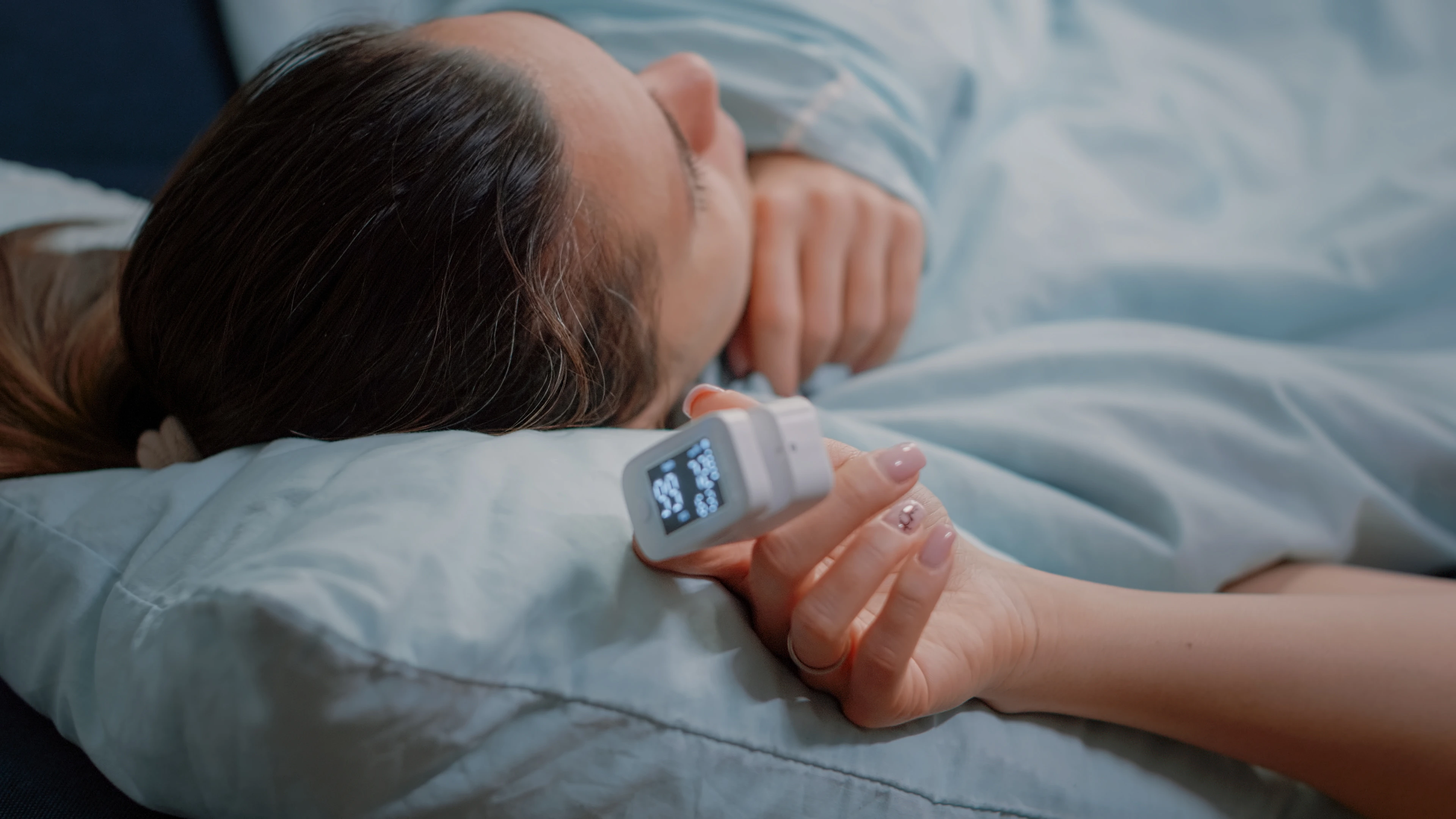
Hypercapnia is a measured blood-gas abnormality, not a diagnosis that symptoms can confirm. Headache, sleepiness, breathlessness, and confusion have many possible causes. A normal fingertip oxygen reading does not rule it out because a pulse oximeter estimates oxygen saturation, not carbon dioxide 34.
How breathing controls carbon dioxide
Cells produce carbon dioxide during metabolism. Blood carries it to the lungs, where it is exhaled. The relevant process is alveolar ventilation, the amount of fresh air reaching the gas-exchanging parts of the lungs each minute. PaCO2 rises when alveolar ventilation falls or cannot increase enough to match carbon dioxide production 2.
A person may be breathing quickly and still ventilating poorly. Severe airway narrowing, shallow breaths, respiratory-muscle fatigue, or a large amount of wasted ventilation can leave too little effective air exchange. This is why respiratory rate alone cannot show whether carbon dioxide clearance is adequate.
Carbon dioxide combines with water in the body and affects acid-base balance. A primary rise in PaCO2 tends to lower blood pH, producing respiratory acidosis. The kidneys respond by retaining more bicarbonate, but that compensation takes time and does not correct the underlying ventilation problem 1.
Acute, chronic, and acute-on-chronic hypercapnia
The same PaCO2 can have different implications depending on how quickly it developed and how much renal compensation is present.
- Acute hypercapnia: PaCO2 rises before the kidneys can retain much bicarbonate. The pH can fall quickly, and neurologic symptoms may be pronounced. An opioid overdose, blocked airway, severe asthma attack, or sudden ventilator problem can cause this pattern.
- Chronic hypercapnia: PaCO2 remains elevated long enough for bicarbonate to rise and bring the pH closer to the normal range. Symptoms may be subtle or absent. Compensation reduces the acidemia; it does not make the underlying ventilatory failure harmless.
- Acute-on-chronic hypercapnia: A person with a chronically elevated PaCO2 develops a further acute rise. The bicarbonate may already be elevated, but the pH falls because the new ventilatory problem outpaces compensation. A COPD exacerbation, infection, sedating drug, mucus plug, or equipment failure can trigger this pattern.
Doctors interpret the full blood gas rather than labeling the pattern from PaCO2 alone. Kidney disease, vomiting, diuretics, sepsis, and other conditions can create mixed acid-base disorders that change the expected pH or bicarbonate 5.
Hypercapnia, hypoxemia, and tissue hypoxia
These terms describe different problems. They can occur together, but one does not prove the others.
| Term | What it means | How it is assessed |
|---|---|---|
| Hypercapnia | Too much carbon dioxide in arterial blood, usually from inadequate effective ventilation | PaCO2 on an arterial blood gas, interpreted with pH and bicarbonate |
| Hypoxemia | Too little oxygen in arterial blood | PaO2 on an arterial blood gas or oxygen saturation measured by co-oximetry; pulse oximetry estimates saturation |
| Tissue hypoxia | Inadequate oxygen available to or used by tissues | Clinical assessment of oxygen delivery, circulation, hemoglobin, organ function, and the cause; there is no single home reading that defines it |
Hypoxemia often contributes to tissue hypoxia, but tissue oxygen delivery also depends on blood flow, hemoglobin, and cellular oxygen use. Hypoxemia can exist without hypercapnia, and hypercapnia may be present while oxygen saturation still looks acceptable 6.
What causes hypercapnia?
Hypercapnia is best understood by asking why effective ventilation has fallen. Several mechanisms can overlap in the same person.
Reduced respiratory drive
The brainstem normally adjusts breathing in response to carbon dioxide and other signals. Opioids, anesthetics, some sedatives, alcohol in combination with other depressants, a brainstem injury, or another central nervous system disorder can reduce that drive. Breaths may become slow, shallow, irregular, or stop. The risk is higher when respiratory-depressing substances are combined or when lung disease, sleep-disordered breathing, or muscle weakness already limits ventilation 1.
Do not stop a prescribed opioid, sedative, or antiseizure medicine abruptly without medical advice. Some medicines can cause dangerous withdrawal if stopped suddenly. A clinician can review the dose, combinations, kidney or liver function, and safer alternatives.
Respiratory-muscle or chest-wall weakness
The diaphragm and other breathing muscles must overcome the load of the lungs and chest wall. Neuromuscular conditions such as myasthenia gravis, Guillain-Barré syndrome, motor neuron disease, muscular dystrophy, or spinal cord injury can weaken that effort. Severe kyphoscoliosis and other chest-wall restrictions can limit breath size. These conditions can cause ventilatory failure without dramatic wheezing or obvious lung damage 2.
Obesity hypoventilation syndrome
Obesity hypoventilation syndrome (OHS) is defined by all three of the following: a body mass index of at least 30 kg/m², sleep-disordered breathing, and an awake resting PaCO2 of at least 45 mm Hg, after other causes of hypoventilation have been excluded 7.
OHS and obstructive sleep apnea (OSA) are related but not interchangeable. Most people with OHS also have OSA, yet OSA by itself does not establish persistent daytime hypercapnia. When an adult with OSA has an elevated awake PaCO2, clinicians look for OHS, COPD-OSA overlap, medication effects, neuromuscular weakness, chest-wall restriction, or another cause rather than assuming OSA alone explains it 7.
Severe airway or lung disease
COPD and a severe asthma exacerbation can increase airway resistance, air trapping, wasted ventilation, and the work of breathing. Hypercapnia appears when the person can no longer maintain enough effective ventilation. Advanced emphysema, severe bronchiectasis, pulmonary edema, extensive pneumonia, or restrictive lung disease can also contribute, particularly when respiratory reserve is already limited. Having one of these diagnoses does not mean a person is always hypercapnic 2.
Upper-airway obstruction and breathing-support problems
A foreign object, swelling, severe upper-airway narrowing, or another obstruction can sharply reduce ventilation. In someone using noninvasive ventilation or a mechanical ventilator, a disconnected circuit, blocked tube, major mask leak, depleted power source, or unsuitable settings can do the same. A new alarm, worsening breathlessness, unusual sleepiness, or failure of prescribed support requires prompt troubleshooting under the person's clinical plan. If breathing is failing, call emergency services rather than trying unprescribed settings.
Rebreathing or high environmental carbon dioxide
Rebreathing exhaled gas in faulty breathing equipment or inhaling a high concentration of carbon dioxide can produce rapid hypercapnia. Occupational or confined-space releases, dry-ice sublimation, beverage-system leaks, and diving or rebreather failures are unusual examples. Carbon dioxide is colorless and odorless, and high concentrations can cause impaired judgment, unconsciousness, convulsions, and death 8.
Do not enter a suspected high-CO2 area to rescue someone unless trained and equipped for the environment. Move away only if it is safe to do so and call emergency services.
Symptoms are not a reliable carbon dioxide test
The effect of hypercapnia depends on the speed and size of the PaCO2 rise, the pH, any accompanying hypoxemia, and the person's baseline. Chronic compensated hypercapnia can cause few symptoms. Acute or worsening respiratory acidosis is more likely to affect the brain 1.
Possible mild or chronic clues include:
- morning headache
- daytime sleepiness or fatigue
- disrupted sleep
- reduced concentration or memory
- breathlessness related to the underlying condition
Acute or severe hypercapnia can cause:
- new confusion, agitation, or unusual behavior
- marked drowsiness or difficulty staying awake
- worsening breathing effort, shallow breathing, or pauses in breathing
- tremor, muscle jerks, or a flapping hand tremor called asterixis
- stupor, seizure, coma, or respiratory arrest
Fever and depressed mood are not useful signs of elevated PaCO2. Flushing, sweating, headache, or a fast heart rate may occur, but they are too nonspecific to diagnose the problem. A person with chronic hypercapnia can also deteriorate without reporting a dramatic sense of breathlessness, especially when respiratory drive or muscle strength is impaired.
When to get emergency help
Call local emergency services for:
- severe or rapidly worsening breathing difficulty, gasping, or inability to speak normally
- blue or gray lips, face, or skin
- inability to wake, extreme sleepiness, new confusion, or collapse
- slow, shallow, irregular, or stopped breathing after a possible opioid or sedative exposure
- a seizure, stupor, or coma
- a blocked airway or suspected high-carbon-dioxide exposure
- failure of prescribed oxygen, PAP, noninvasive ventilation, or mechanical ventilation to support breathing as expected
If opioid overdose is possible, give naloxone if it is available and you know how to use it, then call emergency services. Naloxone can wear off before the opioid does, so apparent improvement does not remove the need for emergency assessment 9.
People who have an emergency oxygen or ventilation plan should follow it while help is coming. Do not withhold emergency oxygen because someone might be at risk of hypercapnia. Clinicians monitor blood gases and adjust oxygen to an appropriate target while treating inadequate ventilation 10.
Arrange a non-emergency medical review for recurring morning headaches, unexplained sleepiness, new exercise limitation, repeated PAP or ventilator alarms, or a chemistry result that raises concern. These findings still need clinical evaluation rather than a home diagnosis.
How doctors diagnose hypercapnia
Arterial blood gas
An arterial blood gas (ABG) directly measures arterial pH, PaCO2, and PaO2. It is the reference test when clinicians need to confirm hypercapnia and assess respiratory acidosis. The pH and bicarbonate help show whether the pattern appears acute, chronic, or mixed. Previous blood gases are valuable because some people with chronic ventilatory failure have a baseline PaCO2 well above the standard reference range 12.
A high PaCO2 does not identify the cause by itself. Doctors combine the result with the breathing pattern, mental status, medication and substance history, lung and neurologic examination, oxygen use, and any respiratory-support data.
A chemistry-panel CO2 result is different
The “CO2” on a routine basic or comprehensive metabolic panel is total carbon dioxide. Most of it is bicarbonate. It is not the same measurement as arterial PaCO2 11.
A high serum bicarbonate can be a clue to chronic respiratory acidosis, but it can also rise for other reasons, including vomiting, diuretics, or a separate metabolic alkalosis. In people with obesity and sleep-disordered breathing whose likelihood of OHS is not high, the American Thoracic Society supports using a serum bicarbonate below 27 mmol/L to help exclude OHS. A value of 27 mmol/L or higher does not diagnose OHS; it indicates that PaCO2 may need confirmation with an ABG in the appropriate clinical setting 7.
Venous blood gas
A venous blood gas can provide useful information about pH and bicarbonate and may help with screening or trends. Venous PCO2 and PO2 are not interchangeable with arterial PaCO2 and PaO2, however. A 2024 systematic review found that venous and arterial differences for PCO2 and PO2 were larger and less predictable than those for pH and bicarbonate 12.
Whether an ABG is still needed depends on the clinical question. It remains important when the exact PaCO2, arterial oxygenation, degree of respiratory failure, or response to ventilatory treatment will affect decisions.
Pulse oximetry and noninvasive carbon dioxide monitoring
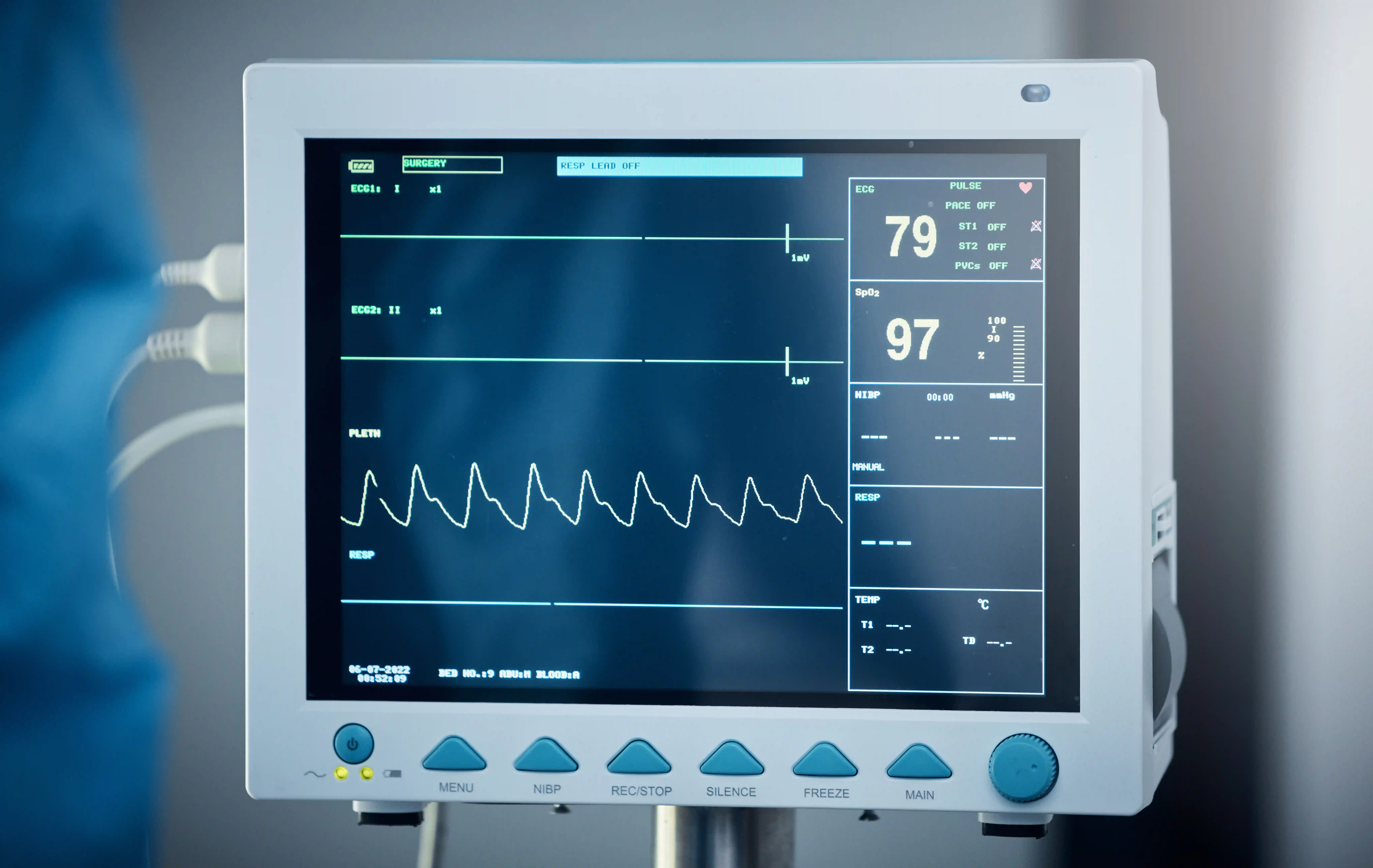
A pulse oximeter estimates peripheral oxygen saturation, or SpO2, and pulse rate. It does not measure PaCO2 or pH. Readings also have accuracy limits, including potential differences associated with skin pigmentation, poor circulation, motion, nail products, and device quality 3.
End-tidal CO2 samples gas at the end of exhalation. Transcutaneous monitoring estimates CO2 through a heated skin sensor. These tools can show trends during anesthesia, emergency care, ventilation, or sleep testing, but neither is automatically identical to PaCO2. Mouth breathing, mask leaks, supplemental oxygen, lung dead space, poor perfusion, and sensor factors can affect the result. Sleep laboratories use end-tidal or transcutaneous CO2 as accepted surrogate measures when assessing sleep hypoventilation, with clinical judgment and calibration checks 13.
Tests that look for the cause
Depending on the situation, evaluation may include:
- medication review, toxicology testing, and neurologic assessment for reduced drive
- chest imaging and lung-function testing for airway, lung, or chest-wall disease
- vital capacity, respiratory-muscle strength, nerve studies, or electromyography when neuromuscular weakness is possible
- a sleep study with appropriate CO2 monitoring when sleep hypoventilation, OHS, or sleep-disordered breathing is suspected
- inspection and data review of PAP, noninvasive ventilation, tracheostomy, or ventilator equipment
Pulmonary-function tests and sleep studies help identify a cause or pattern. They do not replace an ABG when the clinical question is whether awake arterial hypercapnia is present.
How hypercapnia is treated
Treatment depends on the cause, severity, pH, symptoms, and whether the problem is acute or chronic. The immediate objective in dangerous hypercapnia is adequate ventilation and a secure airway, not a fixed PaCO2 number 2.
Acute treatment
Emergency teams may open or protect the airway, support breathing, monitor blood gases, and treat the trigger. Examples include naloxone for suspected opioid toxicity, bronchodilators and anti-inflammatory treatment for severe COPD or asthma exacerbations, antibiotics when a bacterial infection is indicated, removal of an airway obstruction, and correction of a ventilator or circuit problem.
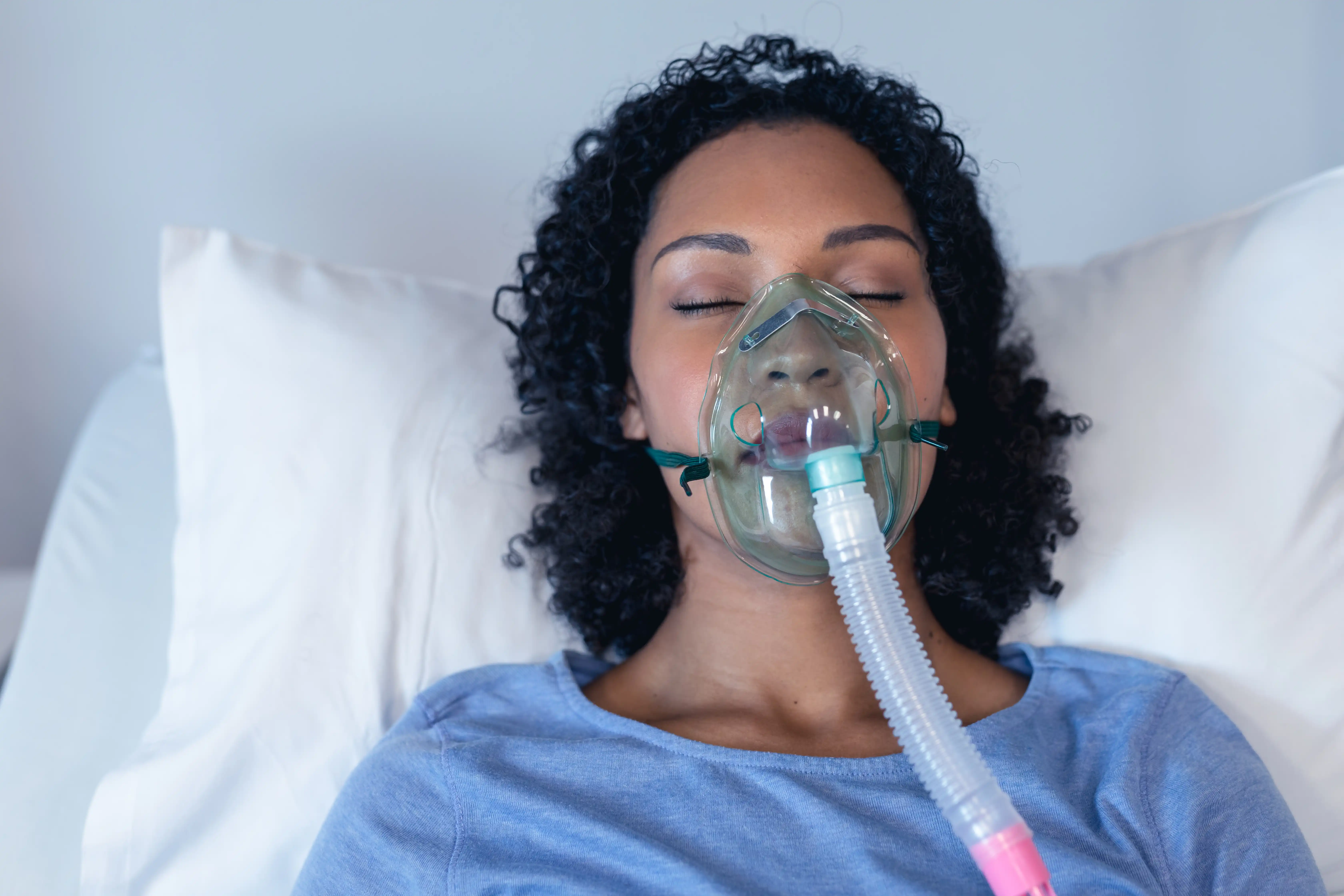
Noninvasive ventilation delivered through a mask can improve alveolar ventilation in selected people. Evidence-based uses include acute or acute-on-chronic respiratory acidosis during a COPD exacerbation and acute ventilatory failure in OHS, provided the person can be monitored and does not need immediate intubation 1415. Invasive mechanical ventilation may be necessary when the airway cannot be protected, breathing stops or remains inadequate, severe instability is present, or noninvasive support is unsuitable or failing.
Oxygen and ventilation do different jobs
Supplemental oxygen treats hypoxemia. It does not, by itself, correct inadequate ventilation or clear carbon dioxide. Some people at risk of hypercapnic respiratory failure need clinician-controlled oxygen and repeat blood gases because excessive oxygen can worsen hypercapnia in certain settings. That is a reason for monitored, targeted treatment, not a reason to deny oxygen during a hypoxemic emergency 10.
Do not change prescribed oxygen flow, CPAP, bilevel PAP, noninvasive ventilation, or ventilator settings unless the treating team has given specific instructions for that situation. Contact the clinical service promptly for new symptoms, repeated alarms, mask or circuit problems, or worsening readings.
Long-term treatment
Chronic care targets the reason ventilation is inadequate:
- OHS and sleep-related hypoventilation: Positive airway pressure is central to treatment. Stable ambulatory OHS with severe OSA is generally treated first with CPAP. Noninvasive ventilation is used in other OHS patterns or when CPAP does not adequately improve symptoms and gas exchange. Sustainable weight-loss treatment may improve or resolve OHS, but it does not replace urgent ventilatory support when respiratory failure is present 7.
- Chronic hypercapnic COPD: Usual COPD treatment remains important. The American Thoracic Society conditionally supports nocturnal noninvasive ventilation in selected people with chronic stable hypercapnic COPD and recommends assessment for OSA before long-term NIV is started 16.
- Neuromuscular and chest-wall disorders: Specialist-led noninvasive ventilation, cough support, secretion management, and monitoring may be needed. Treatment is based on respiratory-muscle function, symptoms, gas exchange, and the course of the underlying condition.
- Medication-related hypoventilation: Prescribers may change the dose, timing, drug combination, or monitoring plan. This should be done with medical supervision rather than abrupt self-discontinuation.
Breathing exercises, posture changes, smoking cessation, and general fitness may support health in selected conditions, but they cannot be relied on to clear dangerous carbon dioxide or reverse respiratory failure.
What not to do
- Do not diagnose hypercapnia from sleepiness, headache, breathlessness, a pulse oximeter, or a chemistry-panel CO2 result.
- Do not adjust oxygen or ventilation settings without the prescribed plan or treating team.
- Do not abruptly stop a prescribed sedative, opioid, or antiseizure medicine without appropriate medical advice.
- Do not breathe into a paper bag. Rebreathing raises inhaled carbon dioxide and can worsen hypoxemia or an unrecognized heart or lung emergency. British Thoracic Society guidance specifically advises against paper-bag rebreathing 10.
- Do not delay emergency help while trying breathing exercises or waiting for a home oxygen number to change.
Frequently asked questions
Can oxygen saturation be normal with high carbon dioxide?
Yes. SpO2 estimates oxygen saturation, not ventilation, PaCO2, or pH. Supplemental oxygen can also keep saturation in range while ventilation remains inadequate. A blood gas or an appropriate clinical CO2 monitor is needed to assess carbon dioxide 4.
Does sleep apnea cause daytime hypercapnia?
Obstructive events can cause temporary CO2 changes during sleep, but OSA alone should not be assumed to explain persistent awake hypercapnia. OHS, COPD-OSA overlap, medicines, neuromuscular weakness, and other causes need consideration. OHS specifically requires awake PaCO2 of at least 45 mm Hg after other causes are excluded 7.
Is a high CO2 value on a metabolic panel hypercapnia?
Not necessarily. That result mainly reflects bicarbonate, while hypercapnia refers to a high partial pressure of carbon dioxide in arterial blood. Serum bicarbonate can support the clinical assessment, but it cannot substitute for PaCO2 11.
Can chronic hypercapnia be safe?
Chronic renal compensation can move pH closer to normal and make symptoms less obvious. It does not show that the cause is harmless or that an acute rise will be tolerated. The person's baseline, pH, symptoms, and underlying disease guide urgency and treatment 1.
Hypercapnia is a sign that ventilation and carbon dioxide production are out of balance. Confirming it requires the right measurement, and treating it requires the cause to be identified. Severe breathing or neurologic changes need emergency care even when a pulse oximeter appears reassuring.