
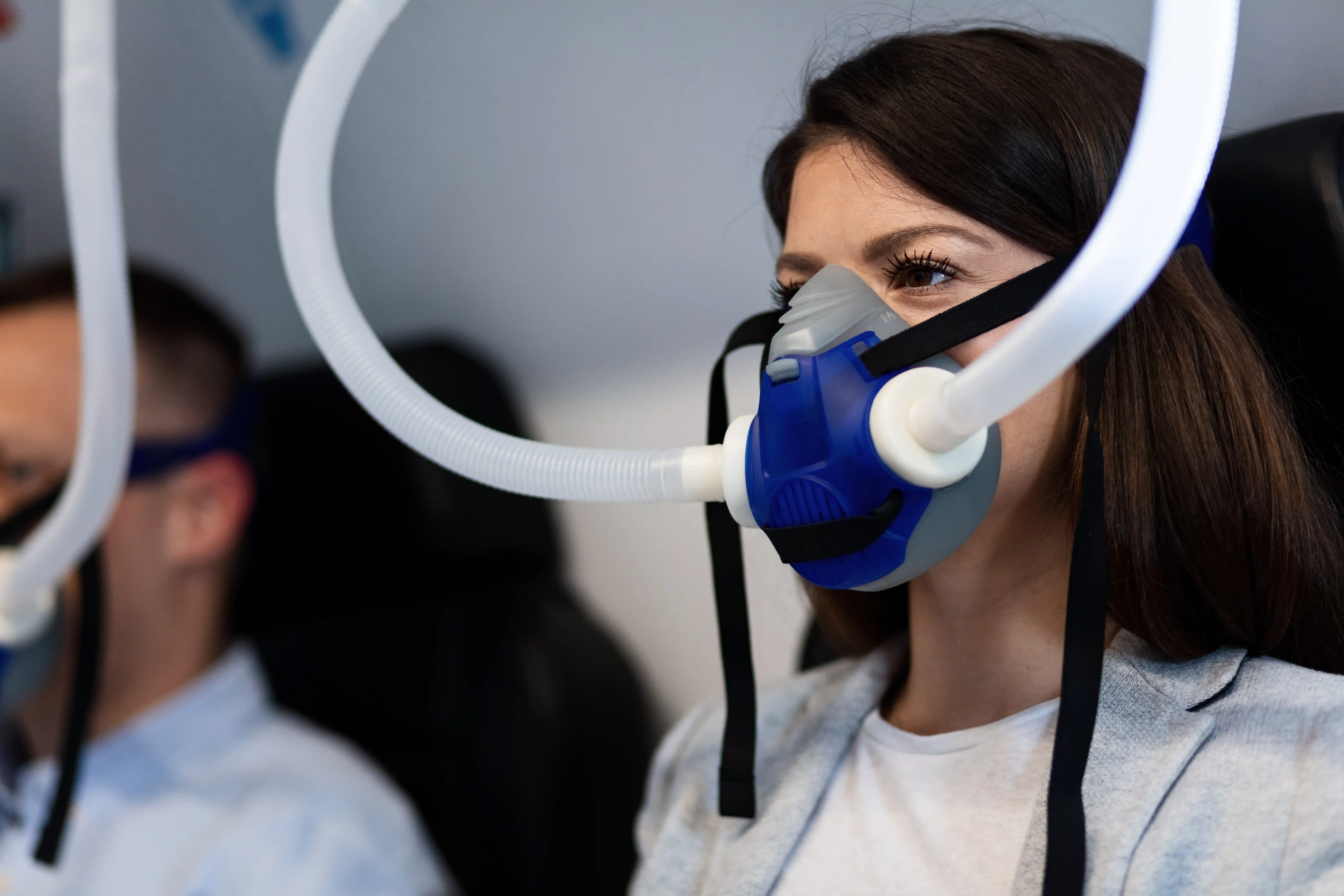
Hypoventilation means the lungs are not moving enough fresh air through the gas-exchanging alveoli to clear the carbon dioxide the body is producing. Breathing may be slow or shallow, but it does not have to look that way. Small ineffective breaths, increased wasted ventilation, weak breathing muscles, a high mechanical load, or reduced brain drive can all raise carbon dioxide even when the breathing rate appears ordinary 1.
Carbon dioxide often rises first during sleep because normal sleep reduces ventilatory drive and muscle activity. Some people therefore have sleep-related hypoventilation while awake carbon dioxide remains normal. Others have chronic awake hypercapnia or develop an acute ventilatory emergency. Symptoms and pulse-oximeter readings cannot determine which pattern is present 1.
Hypoventilation is not the same as low oxygen or sleep apnea
These terms describe different parts of breathing and gas exchange:
| Term | What it describes | Key distinction |
|---|---|---|
| Hypoventilation | Inadequate effective alveolar ventilation relative to carbon-dioxide production | A physiological process that tends to raise arterial carbon dioxide |
| Hypercapnia | An arterial carbon-dioxide level above the expected range | A measured blood-gas finding that can result from hypoventilation |
| Hypoxemia | Too little oxygen in arterial blood | Can occur with or without high carbon dioxide, and can result from problems other than hypoventilation |
| Obstructive apnea | Repeated pauses or major reductions in airflow despite continuing breathing effort | A blocked-airway event; it may coexist with sustained hypoventilation |
| Central apnea | Repeated pauses in airflow without breathing effort during the event | An event pattern, not the same as sustained inadequate ventilation |
| Normal sleep-related change | A modest reduction in ventilation during sleep | Does not by itself meet sleep-hypoventilation scoring criteria 21 |
Hypercapnia is therefore an important consequence and measurement, while hypoventilation is the ventilation problem producing it. Oxygen and carbon dioxide also do not move in perfect lockstep. A person can have low oxygen from lung disease, an apnea, or ventilation-perfusion mismatch without marked carbon-dioxide retention. A person with rising carbon dioxide may initially have a less dramatic oxygen change, especially while receiving supplemental oxygen 13.
Sleep-only, awake, acute, and chronic patterns
During sleep, the brain's response to carbon dioxide becomes less forceful and the contribution of several breathing muscles falls. Rapid eye movement (REM) sleep can expose limited respiratory reserve because most skeletal muscles are less active. Healthy breathing accommodates these changes. In someone with impaired drive, respiratory-muscle weakness, restrictive mechanics, obesity-related load, or severe lung disease, ventilation may become inadequate first during sleep 1.
Sleep-related hypoventilation means carbon dioxide rises abnormally during sleep. Awake blood gases can still be normal. It may precede daytime ventilatory failure in some progressive conditions, but progression is not inevitable and depends on the cause.
Chronic awake hypoventilation causes sustained daytime hypercapnia. Over days to weeks, the kidneys retain bicarbonate to buffer some of the acidity. This compensation can make the condition less obvious, but it does not correct the ventilation problem 1.
Acute hypoventilation develops faster, such as during opioid overdose, severe sedation, a neuromuscular crisis, an acute brainstem problem, or decompensated lung disease. Carbon dioxide can rise before the kidneys have time to compensate, producing respiratory acidosis and potentially rapid deterioration. Acute-on-chronic failure occurs when someone with compensated chronic hypercapnia becomes suddenly worse 1.
What causes hypoventilation?
The useful question is not only whether carbon dioxide is high, but why effective ventilation is inadequate.
Reduced central drive
Opioids, anesthetics, sedatives, and some other medicines or substances can suppress the brain's drive to breathe. Risk can rise with excessive dosing, impaired drug clearance, or combinations of respiratory depressants. Long-term opioid exposure can produce sustained hypoventilation, central apneas, or irregular breathing during sleep, while overdose can cause an acute emergency 1.
Stroke, trauma, inflammation, tumor, surgery, or another disease affecting the brainstem can also impair automatic breathing. The timing and associated neurological signs help determine which investigation is appropriate 1.
Neuromuscular weakness and chest-wall restriction
Motor neuron disease, muscular dystrophy, myopathies, myasthenia gravis, diaphragm dysfunction, spinal cord disease, and other neuromuscular conditions can weaken the pump that moves air. Hypoventilation may appear first during sleep or when lying flat. A weak cough and difficulty clearing secretions add a separate risk.
Severe kyphoscoliosis and other restrictive chest-wall conditions can limit expansion and increase the work required for each breath. These causes are not simply slow-breathing disorders; the respiratory system may be working hard but still fail to generate enough effective ventilation 1.
Severe lung or airway disease
Severe chronic obstructive pulmonary disease (COPD), advanced airway disease, extensive parenchymal disease, or a large increase in physiological dead space can overwhelm respiratory-muscle capacity. Many people with lung disease have hypoxemia without hypercapnia, so the diagnosis still requires carbon-dioxide assessment rather than being inferred from the lung diagnosis or oxygen level 1.
Obesity hypoventilation syndrome
Obesity hypoventilation syndrome (OHS) is not obesity plus tiredness or snoring. It requires obesity, sleep-disordered breathing, and awake arterial carbon dioxide of at least 45 mm Hg after other causes of hypoventilation have been excluded. Obstructive sleep apnea is common in OHS, but OSA alone is not OHS 4.
Obesity can increase respiratory load and reduce lung volume, while sleep-disordered breathing and altered ventilatory control can contribute. Weight is therefore part of the mechanism in OHS, not a complete explanation for every person with obesity and breathing symptoms.
Congenital and other central disorders
Congenital central hypoventilation syndrome (CCHS) is a rare disorder of autonomic breathing control caused by a disease-associated variant in the PHOX2B gene. It often presents in infancy but can be recognized later. Diagnosis requires appropriate clinical findings and genetic testing, not simply unexplained sleep hypoventilation 5.
The International Classification of Sleep Disorders groups sleep-related hypoventilation into six specific categories:
| ICSD category | What the label requires |
|---|---|
| Obesity hypoventilation syndrome | Obesity, sleep-disordered breathing, awake hypercapnia, and exclusion of another main cause |
| Congenital central alveolar hypoventilation syndrome | A compatible autonomic hypoventilation disorder associated with PHOX2B |
| Late-onset central hypoventilation with hypothalamic dysfunction | A rare syndrome with a defined cluster of hypothalamic, behavioral, and autonomic findings after early childhood |
| Idiopathic central alveolar hypoventilation | Central hypoventilation only after pulmonary, neurological, neuromuscular, chest-wall, obesity-related, medication, and congenital causes are excluded |
| Sleep-related hypoventilation due to a medication or substance | A medication or substance is judged to be the cause |
| Sleep-related hypoventilation due to a medical disorder | A pulmonary, neurological, neuromuscular, chest-wall, or other medical disorder is judged to be the cause 1 |
These are diagnostic categories, not labels to assign from symptoms. Terms such as “chronic hypoventilation syndrome” or “central hypoventilation syndrome” without a defined cause can hide important differences in testing and treatment.
Symptoms cannot confirm hypoventilation
Morning headaches, unrefreshing sleep, daytime sleepiness, fatigue, poor concentration, breathlessness with activity, difficulty breathing when flat, and repeated nighttime waking can occur with hypoventilation. A person with neuromuscular weakness may also develop a softer voice, weak cough, trouble clearing mucus, or recurrent chest infections. Chronic hypoxemia or heart strain may contribute to leg swelling.
None of these findings is specific. Sleep apnea, insufficient sleep, medication effects, anemia, heart disease, lung disease, and many other conditions can produce similar symptoms. Some people with significant sleep-related hypoventilation report few symptoms 1.
Severe or rapidly rising carbon dioxide can cause marked drowsiness, confusion, reduced responsiveness, headache, and eventually coma. Blue or gray lips or skin indicate an oxygenation problem and are an emergency sign, but their absence does not rule out dangerous carbon-dioxide retention 1.
How hypoventilation is evaluated
Evaluation needs to establish whether carbon dioxide is high, when it rises, and which mechanism is responsible.
Arterial blood gas and serum bicarbonate answer different questions
An arterial blood gas (ABG) directly measures arterial carbon dioxide and oxygen and shows whether the blood is acidemic. The pH and bicarbonate help distinguish an uncompensated acute rise from a more compensated chronic pattern. One awake ABG does not rule out a sleep-only problem 1.
A standard chemistry panel reports total carbon dioxide, which mostly reflects bicarbonate. An elevated result can be a clue to chronic carbon-dioxide retention, but vomiting, diuretics, and other acid-base conditions can also raise it. In adults with obesity and sleep-disordered breathing whose pretest probability of OHS is low or moderate, the ATS guideline uses a serum bicarbonate below 27 mmol/L to help exclude OHS. That threshold is not a universal hypoventilation screen. When OHS suspicion is high, the guideline recommends measuring an arterial blood gas 4.
A peripheral venous blood gas may help with an initial pH assessment, but venous carbon dioxide varies too much from arterial carbon dioxide to be treated as the same precise value. Venous oxygen cannot substitute for arterial oxygen. Clinicians may use venous results for triage in a specific setting, then obtain an arterial or validated alternative measurement when the exact gas values affect diagnosis or treatment 6.
Pulse oximetry does not measure ventilation
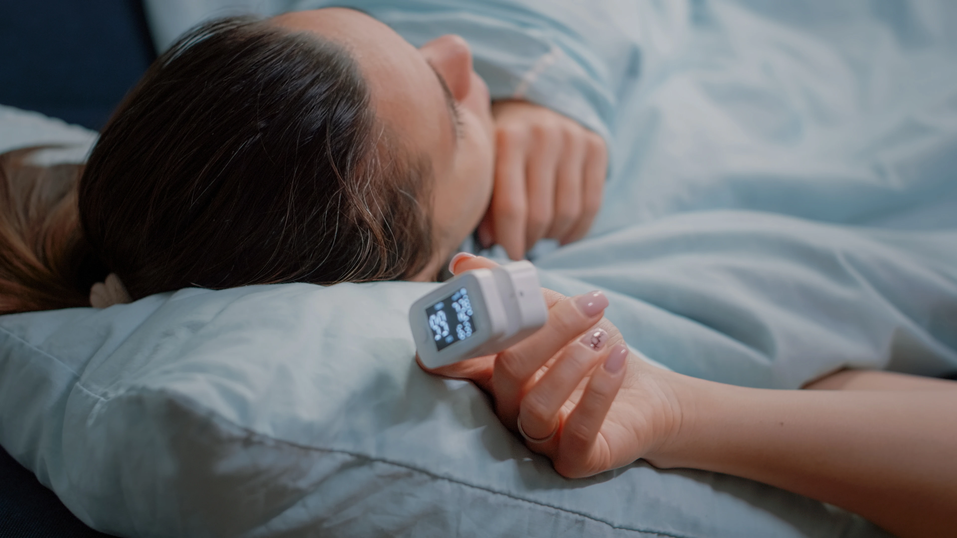
A pulse oximeter estimates hemoglobin oxygen saturation. It does not measure carbon dioxide, breathing effort, or effective alveolar ventilation. Overnight oximetry can identify desaturation that warrants evaluation, but it cannot determine whether the cause is hypoventilation, apnea, lung disease, or another problem. Supplemental oxygen can also improve the displayed saturation while carbon dioxide remains high or continues rising 23.
Sleep testing needs a carbon-dioxide signal
Polysomnography can show sleep stage, airflow, effort, apnea, oxygen saturation, and arousals. To assess sleep-related hypoventilation, the study also needs arterial carbon dioxide or an appropriate surrogate, usually transcutaneous or end-tidal carbon dioxide. These signals have different response times and technical limitations, so clinicians interpret their trends with the rest of the study rather than treating one isolated value as definitive 2.
AASM scoring rules use the following thresholds:
| Population | Sleep-study scoring rule |
|---|---|
| Adults | Arterial CO2 or a validated surrogate above 55 mm Hg for at least 10 minutes, or a rise of at least 10 mm Hg from the awake supine value to above 50 mm Hg for at least 10 minutes |
| Children | Arterial CO2 or a validated surrogate above 50 mm Hg for more than 25% of total sleep time 2 |
These are laboratory scoring criteria, not household cutoffs and not proof of a specific cause. Pediatric interpretation also requires age-appropriate clinical context.
Cause-finding tests are selected from the history
Pulmonary function testing may include spirometry, lung volumes, diffusing capacity, upright and supine vital capacity, or respiratory-muscle pressure measurements. Peak cough flow and airway-clearance assessment can matter in neuromuscular disease. Chest imaging, brain or spinal imaging, cardiac assessment, medication review, toxicology, and selected laboratory tests answer different suspected causes 17.
PHOX2B testing is appropriate when CCHS is clinically suspected. It is not a general test for adult sleep hypoventilation 5. No single panel can replace a cause-directed respiratory and sleep evaluation.
Treatment must correct the cause and support ventilation
Hypoventilation is not treated with one universal machine or lifestyle plan. The urgency and the type of support depend on the cause, acid-base status, alertness, airway protection, secretion burden, and whether the problem is sleep-only, chronic while awake, or acute.
Acute hypoventilation
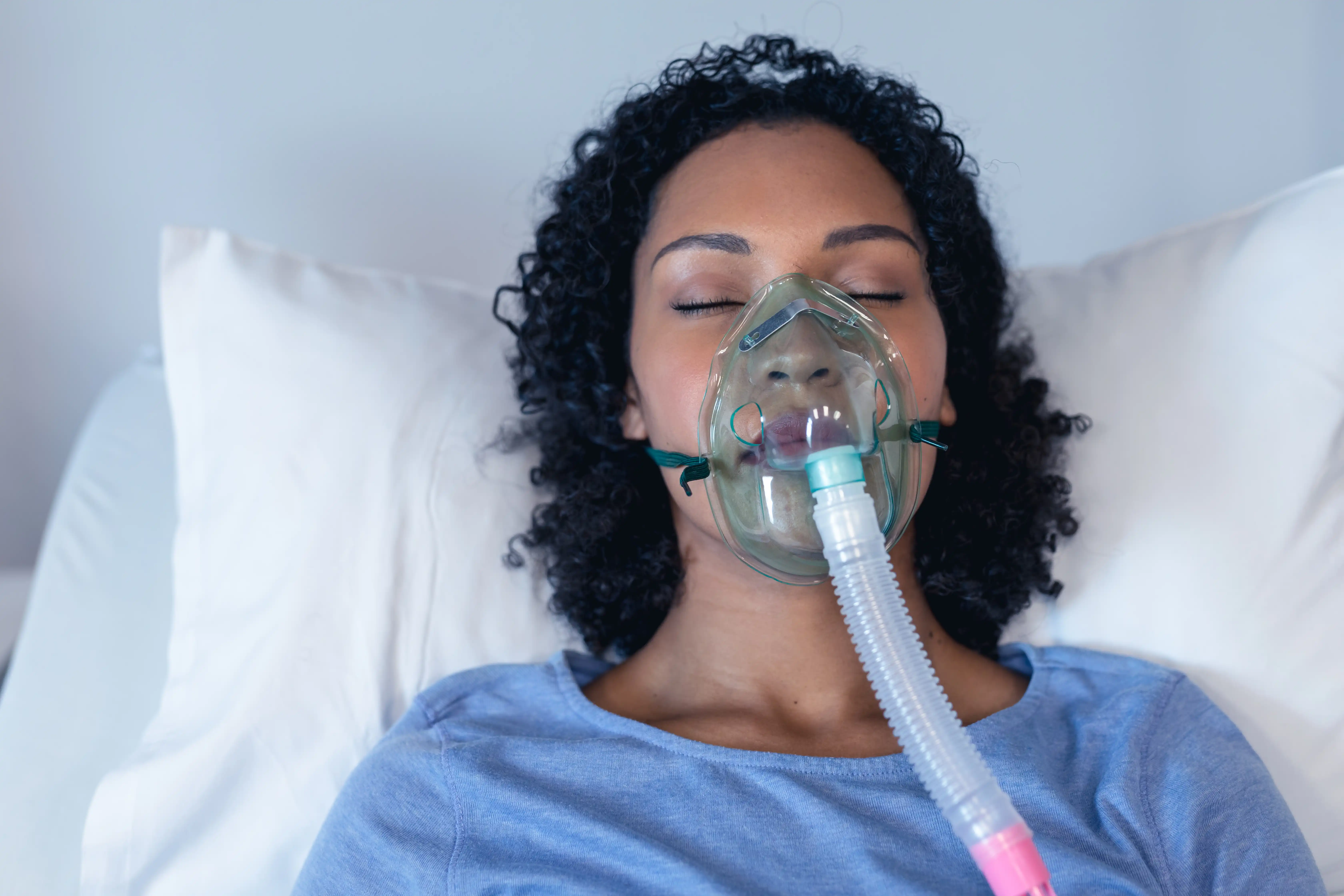
Acute care begins with airway and breathing support while the cause is treated. Selected patients with acute hypercapnic respiratory failure can receive noninvasive ventilation through a mask. Someone who cannot protect the airway, is not breathing adequately, cannot tolerate the interface, or is deteriorating may require invasive ventilation. These decisions require monitored medical care 8.
If opioid overdose is possible, call emergency services and give naloxone if it is available, following the product instructions. Provide rescue breathing or CPR if directed and trained, and remain with the person. Naloxone can reverse opioid effects, but emergency assessment is still necessary because breathing can worsen again 9.
CPAP and noninvasive ventilation do different jobs
Continuous positive airway pressure (CPAP) provides one pressure that splints the upper airway. It does not directly deliver pressure support for each breath. In stable ambulatory OHS with coexisting severe OSA, treating the obstruction with CPAP is the recommended first approach for many patients 4.
Noninvasive ventilation (NIV), often delivered with bilevel pressures and sometimes a backup rate, can augment ventilation. It is used in selected people with OHS without severe OSA, persistent hypoventilation despite an appropriate CPAP trial, neuromuscular weakness, restrictive chest-wall disease, central hypoventilation, or other defined ventilatory failure. Mode, interface, pressures, backup rate, and oxygen are prescribed and checked against symptoms, carbon dioxide, oxygenation, and device data 10.
Do not switch between CPAP and bilevel treatment or change pressure and backup settings without the treating respiratory or sleep team. A mask feeling stronger does not show that ventilation is safe or effective.
Cause-specific long-term care
OHS care includes prescribed PAP and a medically supported plan for sustained weight reduction. Weight treatment can reduce respiratory load over time, but it does not replace PAP, gas monitoring, or acute respiratory care 4.
Neuromuscular care may combine NIV with assisted cough, mechanical insufflation-exsufflation, secretion management, and treatment of swallowing or aspiration risk. Airway clearance and ventilation are related but separate needs 7.
Chest-wall, central-drive, and congenital disorders may require scheduled ventilatory support during all sleep and, in more severe cases, while awake. People with CCHS need specialist plans based on their PHOX2B variant, awake and asleep physiology, and other autonomic risks 5.
Severe lung or airway disease needs condition-specific treatment, such as relieving airflow obstruction, treating an exacerbation or infection, and managing secretions. Long-term NIV is appropriate only for selected hypercapnic lung-disease populations, not for every low oxygen reading or breathless patient 11.
Medication-related hypoventilation requires a clinician-led review of dose, interactions, organ function, and alternatives. Do not abruptly stop a prescribed opioid, benzodiazepine, or other dependence-forming medicine unless emergency clinicians direct it.
Oxygen treats oxygenation, not ventilation
Supplemental oxygen can treat hypoxemia, but it does not remove carbon dioxide or restore an ineffective breath. Some people need oxygen in addition to ventilatory support. In people at risk of hypercapnic respiratory failure, the oxygen target and delivery method need a plan plus blood-gas or carbon-dioxide reassessment because excessive oxygen can worsen carbon-dioxide retention in susceptible conditions 3.
Do not start, stop, or increase home oxygen based only on symptoms or a consumer pulse oximeter. A normal saturation on oxygen does not prove that ventilation is adequate.
When to get urgent help
Call emergency services for severe or rapidly worsening breathing difficulty, blue or gray lips or skin, new confusion, inability to stay awake or wake normally, gasping or irregular breathing, a seizure, or suspected medication or drug overdose. If opioid overdose is possible, use naloxone if available while emergency help is coming 9.
A person with a neuromuscular condition needs prompt respiratory assessment for a newly weak cough, trouble clearing secretions, choking, difficulty lying flat because of breathing, rapidly declining strength, morning headaches, or new daytime sleepiness. Waiting for a low pulse-oximeter number can miss early ventilatory failure 7.
Do not drive or perform hazardous work if sleepiness, confusion, or impaired alertness makes it difficult to stay awake. Arrange another way to travel and discuss return-to-driving safety with the treating clinician 12.
Common questions
Can a home pulse oximeter detect hypoventilation?
No. It can identify low estimated oxygen saturation, but it cannot measure carbon dioxide or distinguish hypoventilation from apnea, lung disease, or a technical artifact. Suspected sleep hypoventilation needs appropriate carbon-dioxide assessment.
Is hypoventilation the same as sleep apnea?
No. Apneas are repeated airflow events. Hypoventilation is sustained inadequate effective ventilation that raises carbon dioxide. Obstructive or central apneas can coexist with hypoventilation, so a sleep study may need to measure both event patterns and carbon dioxide.
Can oxygen correct hypoventilation?
Oxygen can correct low oxygen in some situations, but it does not provide ventilation or clear carbon dioxide. When carbon-dioxide retention is possible, oxygen should be prescribed as part of a monitored plan rather than used alone as a substitute for ventilatory support.